On Heart Kardiohirurgija.rs
Using our unlabeled heart diagrams, you can challenge yourself to identify the individual parts of the heart as indicated by the arrows and fill-in-the-blank spaces. This exercise will help you to identify your weak spots, so you'll know which heart structures you need to spend more time studying with our heart quizzes.

Cardiovascular Disease
In animals with lungs —amphibians, reptiles, birds, and mammals—the heart shows various stages of evolution from a single to a double pump that circulates blood (1) to the lungs and (2) to the body as a whole. In humans and other mammals and in birds, the heart is a four-chambered double pump that is the centre of the circulatory system.

Heart Anatomy Labeled Diagram, Structures, Blood Flow, Function of Cardiac System — EZmed
Anatomy The heart is an organ that weighs approximately 350 grams (less than one pound). It's nearly the size of an adult's clenched fist. It's located in the thorax (chest)—between the lungs —and extends downward between the second and fifth intercostal (between the ribs).

Anatomy and Physiology Heart Anatomy
The structure of the heart If you clench your hand into a fist, this is approximately the same size as your heart. It is located in the middle of the chest and slightly towards the left. The.
Structure and Function of the Heart
Clinical notes Sources Related articles + Show all Heart anatomy The heart has five surfaces: base (posterior), diaphragmatic (inferior), sternocostal (anterior), and left and right pulmonary surfaces. It also has several margins: right, left, superior, and inferior:

External Structure Of Heart Anatomy Diagram
$9.99 Add To Cart Anatomy of the Heart Welcome to the anatomy of the heart made easy! We will use labeled diagrams and pictures to learn the main cardiac structures and related vascular system. In addition to reviewing the human heart anatomy, we will also discuss the function and order in which blood flows through the heart.

12+ Human Heart Location Diagram Robhosking Diagram
What does a heart diagram look like? The inside and outside of your heart contain components that direct blood flow: Inside of the Heart Outside of the Heart Function What is the heart's function? Your heart's main function is to move blood throughout your body. Your heart also: Controls the rhythm and speed of your heart rate.

Image Of The Heart Labeled
The myocardium is functionally the main constituent of the heart and the thickest layer of all three heart layers. It is a muscle layer that enables heart contractions. Histologically, the myocardium is comprised of cardiomyocytes.Cardiomyocytes have a single nucleus in the center of the cell, which helps to distinguish them from skeletal muscle cells that have multiple nuclei dispersed in the.

Heart anatomy, Heart diagram, Human heart diagram
The user can show or hide the anatomical labels which provide a useful tool to create illustrations perfectly adapted for teaching. Anatomy of the heart: anatomical illustrations and structures, 3D model and photographs of dissection Illustrated anatomy of the heart: illustrations and photography

How to Draw the Internal Structure of the Heart 13 Steps
What are the different coronary arteries? The 2 main coronary arteries are the left main and right coronary arteries. Left main coronary artery (LMCA). The left main coronary artery supplies blood to the left side of the heart muscle (the left ventricle and left atrium). The left main coronary divides into branches:
Heart Diagram Labeled
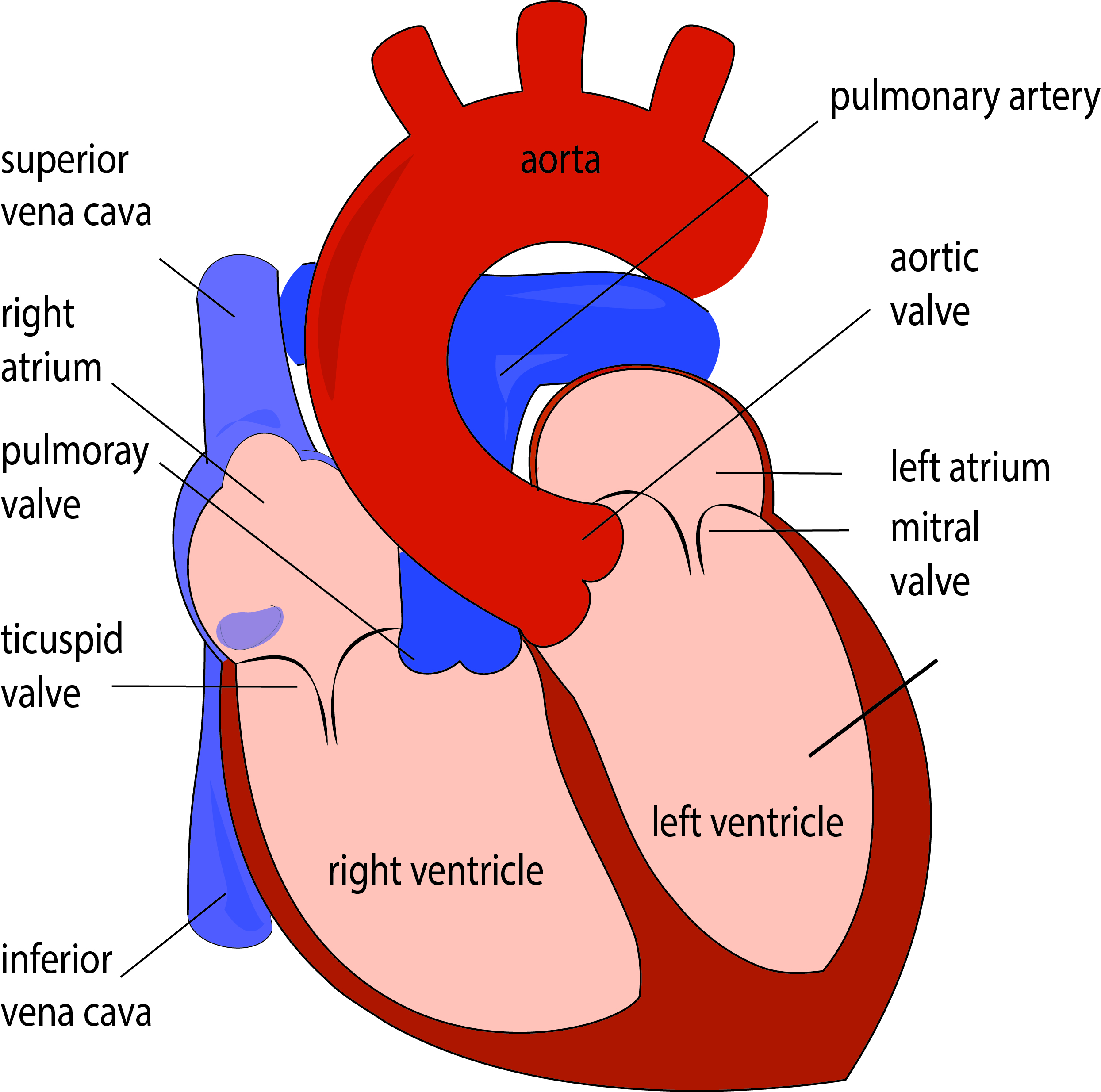
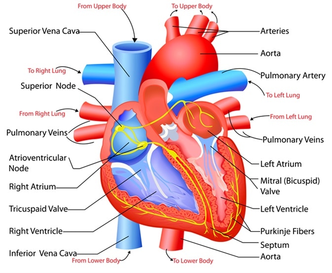
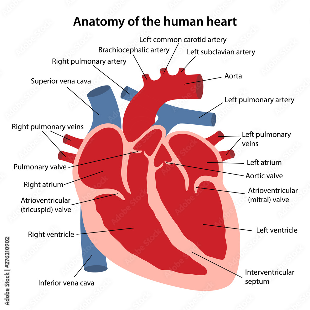
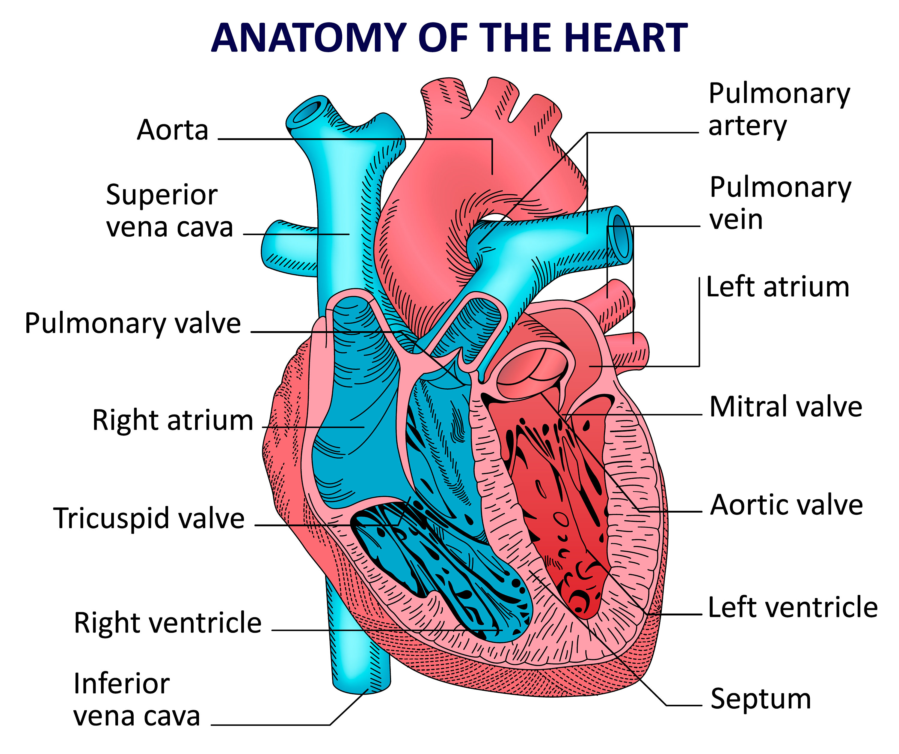
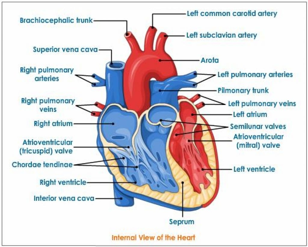
heart diagram, Labeled correctly Contents. 1 Summary. 1.1 SVG; 1.2 PNG, JPG. English: Diagram of the human heart. 1. Superior vena cava 2. 4. Mitral valve 5. Aortic valve 6. Left ventricle 7. Right ventricle 8. Left atrium 9. Right atrium 10. Aorta 11. Pulmonary valve 12. Tricuspid valve. 13. Inferior vena cava
Human heart anatomy. Vector diagram Etsy
The human heart is located within the thoracic cavity, medially between the lungs in the space known as the mediastinum. Figure 19.2 shows the position of the heart within the thoracic cavity. Within the mediastinum, the heart is separated from the other mediastinal structures by a tough membrane known as the pericardium, or pericardial sac.

Heart Anatomy Anatomy and Physiology II
In this interactive, you can label parts of the human heart. and drop the text labels onto the boxes next to the heart diagram. If you want to redo an answer, click on the box and the answer will go back to the top so you can move it to another box. If you want to check your answers, use the Reset Incorrect button.
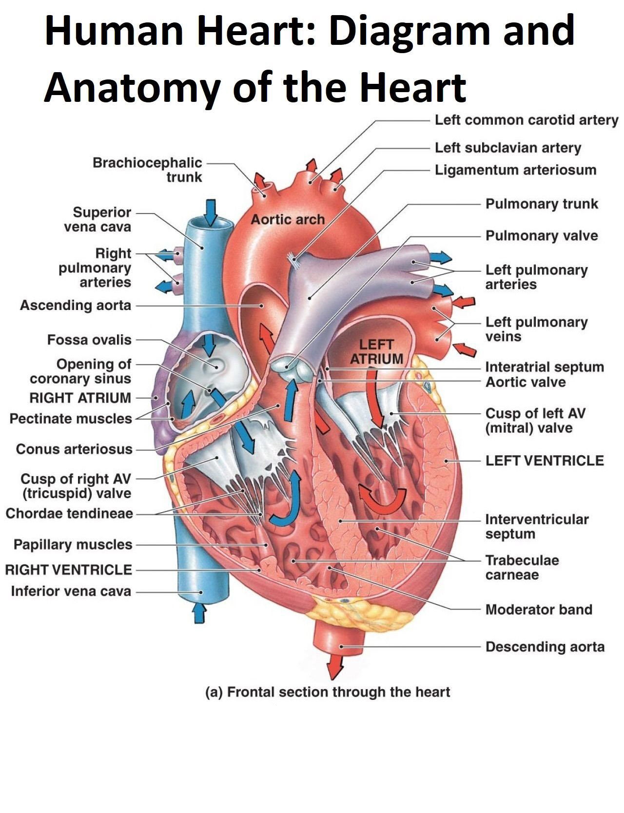
Diagram and Anatomy of the Heart Poster Etsy
A typical heart has two upper and two lower chambers. The upper chambers, the right and left atria, receive incoming blood. The lower chambers, the more muscular right and left ventricles, pump blood out of the heart. The heart valves, which keep blood flowing in the right direction, are gates at the chamber openings.

15 Heart Diagram Labeled Blood Flow Robhosking Diagram
Discussed in this video is how to draw and label the structures of the heart, the layers of the heart, and a discussion on how blood flows within the heart.
31 Label The Heart Diagram Label Design Ideas 2020
The heart is made of three layers of tissue. Endocardium is the thin inner lining of the heart chambers and also forms the surface of the valves.; Myocardium is the thick middle layer of muscle that allows your heart chambers to contract and relax to pump blood to your body.; Pericardium is the sac that surrounds your heart. Made of thin layers of tissue, it holds the heart in place and.